Biomechanics
Finite Element Based Modeling of Keratorefractive Surgeries
Approximately one-fourth of the world’s population suffers from nearsightedness. Since the shape of the cornea greatly influences the refractive power, an opthalmologist may reshape the cornea by surgery in order for light rays to naturally focus on the retina. Radial keratotomy (RK) was one of the original procedures. Correction was achieved by flattening the central cornea for nearsightedness (myopia) for a more uniform corneal curvature. A refractive error means that the anterior surface of the eye does not bend light correctly on the retina, resulting in a blurred image. Clear vision is a result of light rays passing through the cornea, pupil, and lens and focusing directly upon the retina. If the geometrical shape of the cornea is too steep or too flat in relation to the shape of the eye, light rays focus either in front of or behind the retina resulting in refractive errors, such as nearsightedness, farsightedness, or astigmatism. Laser Assisted In-Situ Keratomileusis (LASIK) is a popular surgical alternative for the correction of myopia and other eye conditions. In this technique, a hinged flap is created and folded back, and the exposed stroma is photoablated using an excimer laser.
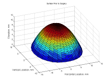
The finite element method is a numerical procedure for analysis of structures using simultaneous algebraic equations based on the degrees of freedom (displacement in stress analysis, temperatures in heat transfer). In this research, we use a finite element based model to simulate the response of a human eye to Lasik procedure. The cornea is modeled as a thick membrane shell. The topography of a patient’s cornea is measured using a corneal mapping device, and used to generate the finite element mesh. Additional model parameters include patient specific data such as the corneal thickness distribution, constitutive response, and intraocular pressure distribution. The sensitivity of the proposed model to the surgical parameters described has been studied. The expected curvature distribution is compared with the clinically obtained post-op curvature distribution.
Schematics of the LASIK procedure, a flap is made and the laser is applied to the anterior region of the cornea
C. Deenadayalu, B. Mobasher, S. D. Rajan and G. Hall “Refractive Change Induced by the Lasik Flap in a Biomechanical Finite Element Analysis Model”, Journal of Refractive Surgery, 22:3, 1-7, 2006. [download]

