Imaging Technologies
Photoacoustic Endoscopy
Photoacoustic endoscopy (PAE) has emerged as a minimally invasive tool for imaging internal organs and tissues. One of the main themes of our work involves the first reported dual-intrauterine photoacoustic and ultrasound deep-tissue imaging endoscope. This device was designed to enable physicians, at the point-of-care, to better elucidate overall gynecological health by imaging the lining of the human uterus. In order to further miniaturize the diameter of our endoscopes alternative PAE designs were investigated utilizing hollow optical waveguides, in order to allow for con-centric guiding of both light and sound.
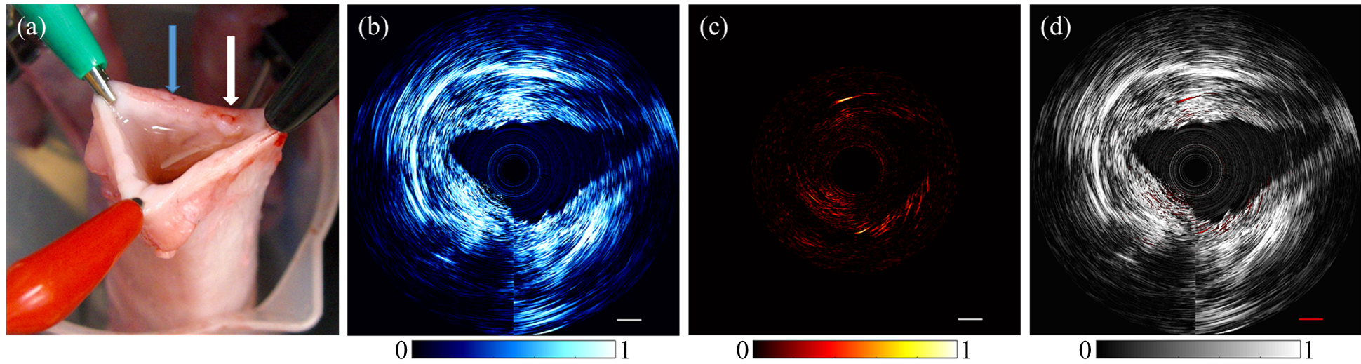 Figure: (a) Photograph of an excised adult pig uterus. Blue arrow indicates the location of blood injection. White arrow indicates location of water injection. (b)Ultrasound reconstruction of pig uterus. (c) Photoacoustic reconstructions of same axial position as (b). (d) Overlap of photoacoustic data on ultrasound data. Scale bar represents 3 mm 1.
Christopher Miranda, Joel Barkley, and Barbara S. Smith* “Intrauterine Photoacoustic and Ultrasound Imaging Probe.” Journal of Biomedical Optics. 23.4 (2018): 046008.
Waveguides
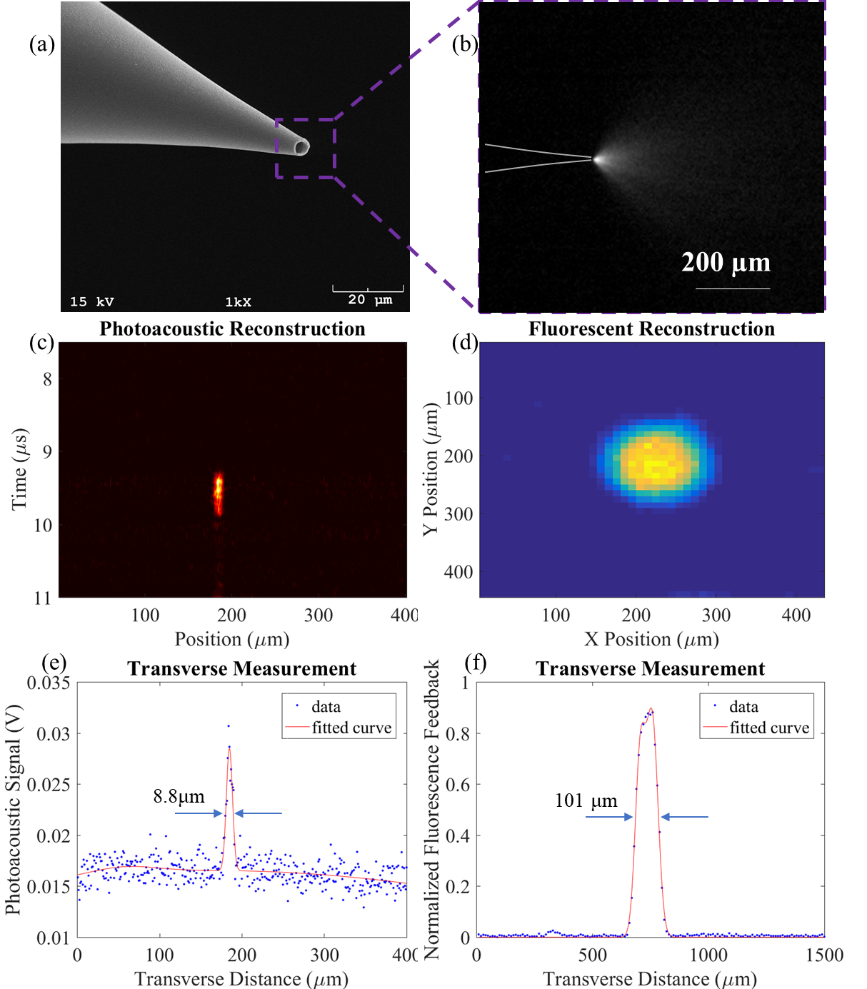
Our waveguide technology has been further expanded for use in micropipette electrodes, common in the field of single cell electrophysiology. By coupling excitation light to these micropipettes, photoacoustic and fluorescence feedback can be generated from select neurons. This enables targeted robotic automation for high resolution recording of neuronal activity in healthy and diseased states.